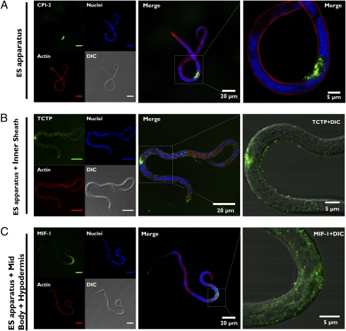
Fig. 3.
Immunolocalization studies of ES proteins in B.malayi mf reveal three different expression patterns. (A) Protein localization exclusively in the ES apparatus. Representative staining is shown for CPI-2. Also exhibiting the same pattern, VAL-1, TPI, and a fraction of mf stained with anti-TCTP (Fig. S3). (B) Protein localization in either the ES apparatus or the mf inner sheath. This pattern was exhibited in another fraction of specimens stained for TCTP. (C) The third pattern, observed for MIF-1, consisted of prominent expression through the midbody. Images on the Right of B and C are presented as merged images of single planes showing signal presence in the ES-vesicle and differences in compartmentalization of TCTP and MIF-1 toward the parasite surface.

