Fig. 2.
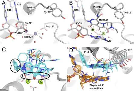
Active site region of the PFV intasome bound to metal cofactors and INSTIs. With the exception of carbon atoms, standard atom coloring is used throughout: oxygen, red; nitrogen, blue; sulfur, yellow; fluorine, pale blue; chlorine, green; manganese, purple; magnesium, light green; phosphorus, orange. (A) The intasome active site in Mn2+-bound form. Protein is represented as a gray cartoon. Stick representations are shown for side chains mentioned in the text and DNA, colored by atom with gray used for carbon. Large purple spheres indicate Mn atoms and smaller red spheres show the water molecules coordinated to the metal atoms. (B) The Mg2+/MK2048-bound intasome active site with viewpoint and representation as in A. Here, MK2048 is also shown as a stick representation and Mg atoms as spheres. (C) Comparison of the different inhibitor-bound structures. Inhibitor molecules are shown as sticks with cyan carbon atoms. The black ovals highlight the conserved features of these drugs: 1, the metal chelating heteroatoms; and 2, the halogenated benzene ring. (D) An alternative view comparing drug binding, rotated 90° about the horizontal axis compared with C. Here, the side chain of Tyr212 is shown to highlight its involvement in binding different INSTIs; DNA is shown as sticks with carbon atoms colored orange.

