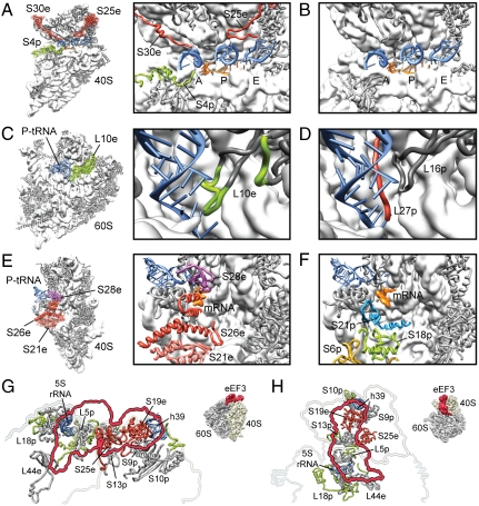
Fig. 3.
Functional roles for eukaryote-specific r proteins. (A) Small 40S subunit with newly modeled r-proteins S30e and S25e (red) and eukaryote-specific extension of S4p (green) highlighted (thumbnail, Left; zoom, Right). (B) Comparative view of the bacterial 30S subunit decoding site (11, 12). In A and B, the anticodon-stemloops of A-, P- and E-tRNAs (blue) and mRNA (orange) are shown for reference. (C) Large 60S subunit with eukaryote-specific extension of L10e (green) highlighted (thumbnail, Left; zoom, Right). (D) Comparative view of the bacterial 50S subunit with bacterial-specific L27p colored red (11). In C and D, the acceptor-stem of the P-tRNA (blue) is shown for reference. (E) Small 40S subunit with newly modeled r-proteins S21e, S26e, and S28e colored distinctly (thumbnail, Left; zoom, Right). (F) Comparative view of the bacterial 30S subunit with bacterial-specific S18p shown in green (11). In E and F, the P-tRNA (blue) and mRNA (orange) are shown for reference. (G and H) The binding site of eEF3 on the S. cerevisiae 80S ribosome, with (G) side and (H) top views (see insets) showing the binding site of eEF3 as a red outline and molecular models of ribosomal components that comprise the eEF3 binding site. Newly identified proteins are shown in red (S19e, S25e) and newly modeled r-protein extensions in green, whereas core r proteins are colored gray. Modified from ref. 48.

