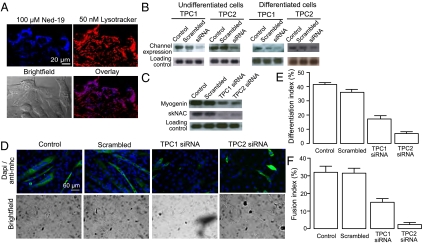
Fig. 7.
C2C12 differentiation is altered by down-regulation of TPCs on acidic organelles. (A) Confocal microscopy of undifferentiated C2C12 cells. (Upper Left) Labeling of NAADP receptor with 100 μM Ned-19. (Upper Right) Labeling of acidic organelles with 50 nM lysotracker. (Lower Left) Bright field image of C2C12 cells. (Lower Right) Overlay of Ned-19 and lysotracker labeling showing colocalization. (B) C2C12 cells were transfected with siRNA against TPC1 or TPC2, respectively. RNA was harvested either 24 h after transfection (undifferentiated cells) or 24 h after differentiation was induced (differentiated cells). The RNA was analyzed for expression of TPC1 or TPC2, respectively, by Northern blot analysis. (C) C2C12 cells were transfected with siRNA against TPC1 or TPC2 for 24 h and differentiated for 24 h. RNA was harvested and analyzed for expression of myogenin and skNAC by Northern blot analysis. (D) C2C12 cells were transfected with siRNA against TPC1 or TPC2 for 24 h and differentiated for 4 d. The cells were stained for myosin heavy chain and DAPI to determine the differentiation index (percent nuclei in myosin heavy chain-positive cells). (E) Bar chart (mean with SEM; n = 4) representing the differentiation index following treatment for 4 d with control, scrambled siRNA, TPC1 siRNA, or TPC2 siRNA. (F) Bar chart (mean with SEM; n = 4) representing the fusion index (percent nuclei in myosin heavy chain-positive cells with at least three nuclei) following treatment for 4 d with control, scrambled siRNA, TPC1 siRNA, or TPC2 siRNA.

