Fig. 5.

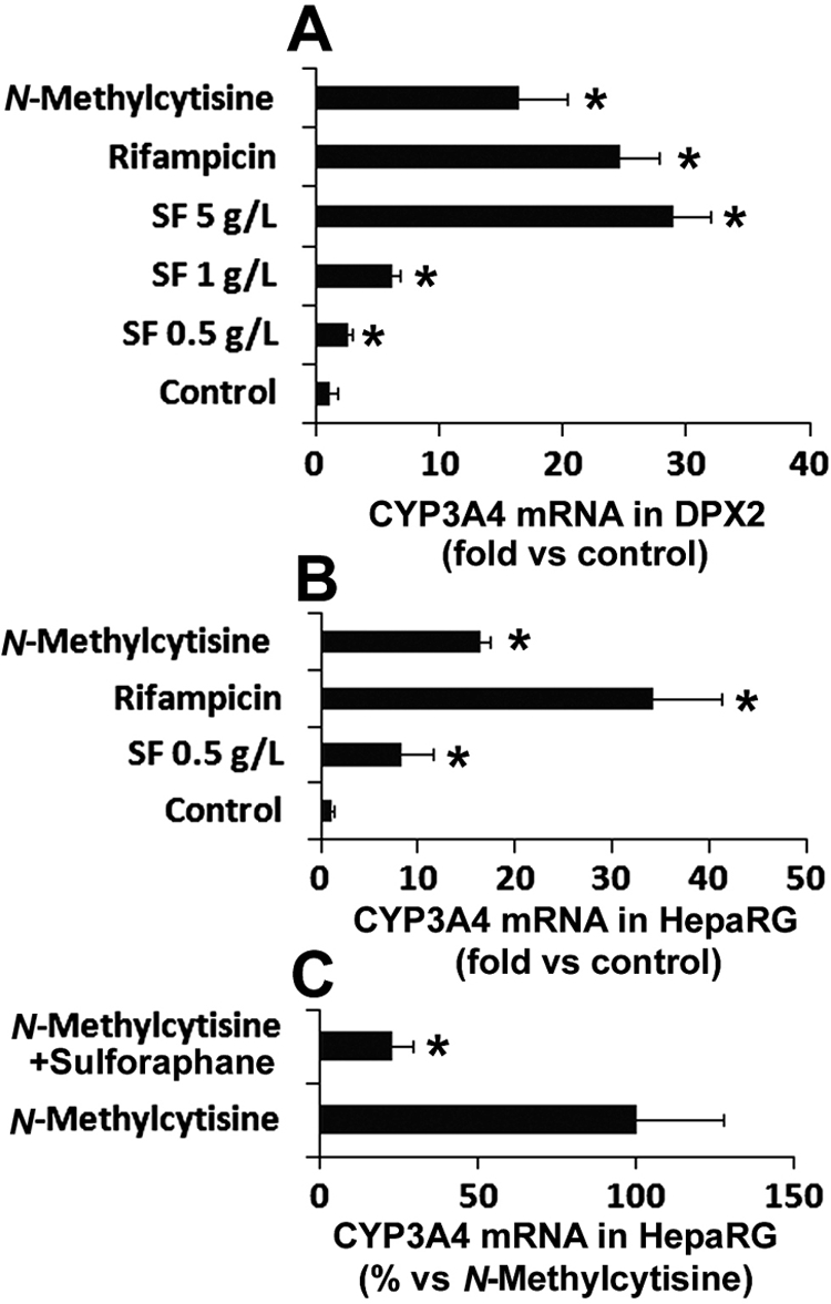
Induction of CYP3A4 by SF aqueous extract and N-methylcytisine. DPX2 cells and HepaRG cells were incubated with SF aqueous extract and N-methylcytisine (10 μM) for 48 h. CYP3A4 mRNA expression was analyzed by qPCR. Values were quantified using the comparative cycle threshold method, and samples were normalized to glyceraldehyde-3-phosphate dehydrogenase. Rifampicin (10 μM) served as a positive control of CYP3A4 inducer. A, effect of SF aqueous extract and N-methylcytisine on CYP3A4 expression in DPX2 cells. The data are shown as the fold induction versus control (n = 3; *, p < 0.05 versus control). B, effect of SF aqueous extract and N-methylcytisine on CYP3A4 expression in HepaRG cells. The data are shown as the fold induction versus control (n = 3; *, p < 0.05 versus control). C, effect of PXR antagonist sulforaphane (20 μM) on N-methylcytisine-mediated CYP3A4 induction in HepaRG cells. CYP3A4 expression in the N-methylcytisine-treated group was set as 100% (n = 3; *, p < 0.05 versus N-methylcytisine-treated group).
