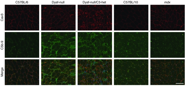
Figure 4. Immunofluorescence analysis of complement C5b-9 deposition in mouse skeletal muscles.
Immunofluorescence analysis did not detect complement C5b-9 expression in the muscle sections from WT mice (C57BL/6 and C57BL/10) and mdx mice but showed that complement C5b-9 was deposited onto the muscle cell membrane in dysferlin-null mice. All samples were prepared and processed equally at the same time, and all images were taken at the same exposure level. Scale bar: 100 μm.

