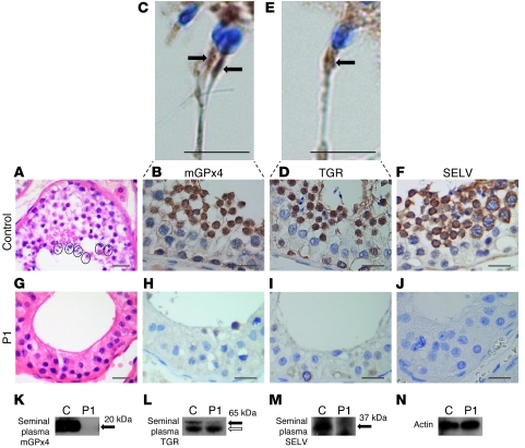
Figure 2. Spermatogenic maturation arrest associated with deficiencies of testis-expressed selenoproteins.
H&E images (original magnification, ×40) of a seminiferous tubule from a normal subject (A) and P1 (G). Circled areas (A) from normal testis show mature spermatozoa. Spermatids and spermatozoa were absent, but with preservation of spermatogonia and spermatocytes, in P1 (G). IHC showed virtual absence of mGPx4, TGR, and SELV immunoreactivity in testis from P1 (H–J) compared with their expression in later stages of spermatogenesis in a normal subject (B, D, and F). Expanded images (C and E; original magnification, ×60) also highlighted mGPx4 and TGR expression in the midpiece of normal spermatozoa (arrows). Scale bars: 20 μm (A, B, D, and F–J); 5 μm (C and E). Western blotting of seminal plasma from a normal control subject and P1 using antibodies for mGPx4 (K), TGR (L), and SELV (M), with actin (N) as a loading control. Black arrows indicate specific bands; white arrow denotes nonspecific band.

