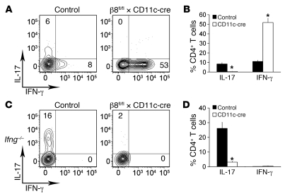
Figure 1. Impaired Th17 development in β8fl/fl × CD11c-cre mice.
(A and C) Flow cytometry plots of control and β8fl/fl × CD11c-cre (A) or Ifng–/– control and Ifng–/– β8fl/fl × CD11c-cre (C) colon lamina propria CD4+ T cells stained for IL-17 and IFN-γ after activation with PMA/ionomycin. Numbers in quadrants indicate the percentage of positive cells. (B and D) Percentage of IL-17– and IFN-γ–producing CD4+ T cells isolated from the colon lamina propria of control and β8fl/fl × CD11c-cre (B) or Ifng–/– control and Ifng–/– β8fl/fl × CD11c-cre (D) mice after activation with PMA/ionomycin. Data represent the mean ± SEM of 3 independent experiments and more than 6 mice per group. *P < 0.005.

