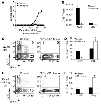
Figure 2. β8fl/fl × CD11c-cre mice are protected from EAE.
(A) Clinical scores of control and β8fl/fl × CD11c-cre mice after EAE immunization. (B) Total number of CD4 and CD8 T cells isolated from the CNS of control or β8fl/fl × CD11c-cre mice on day 19 after EAE immunization. (C) Flow cytometry plots of CD4+ T cells from the CNS of control or β8fl/fl × CD11c-cre mice on day 19 after EAE immunization. Cells were activated with PMA/ionomycin and then stained for IL-17 and IFN-γ. (D) Percentage of IL-17– and IFN-γ–producing CD4+ T cells from the CNS of control and β8fl/fl × CD11c-cre mice. (E) Flow cytometry plots of CD4+ T cells from the draining lymph nodes of control or β8fl/fl × CD11c-cre mice on day 8 after EAE immunization. Cells were activated with PMA/ionomycin and then stained for IL-17 and IFN-γ. (F) Percentage of IL-17– and IFN-γ–producing CD4+ T cells from the draining lymph nodes of control and β8fl/fl × CD11c-cre mice. Data represent the means ± SEM of 3 independent experiments and at least 10 mice in each group. *P < 0.01.

