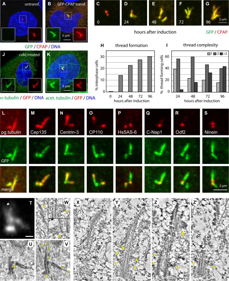
Figure 2. CPAP overexpression induces the formation of abnormal elongated centrioles.
(A-B) U2-OS cells not transfected (A) or transiently transfected with GFP-CPAP for 72 h (B) and stained with antibodies against GFP (green) and CPAP (red).
(C-I) i-GFP-CPAP cells induced with doxycycline for 0, 24, 48, 72 or 96 h prior to fixation and stained with antibodies against GFP (green) and CPAP (red). Representative thread configurations are shown in (C-G). Panel (H) shows the frequency of threads (N>250 at each time point), panel (I) their complexity, as determined by the number of free ends per thread.
(J-K) U2-OS cells transiently transfected with GFP-CPAP for 72 h, incubated for 1 h at 4°C and stained with antibodies against α-tubulin (green) and GFP (red) (J), or with antibodies against acetylated tubulin (green) and GFP (red).
(L-S) Representative high magnification images of threads from i-GFP-CPAP cells at different stages of the cell cycle induced with doxycycline for 48 h and stained with antibodies against GFP (green) and polyglutamylated (pg.) tubulin (L), Cep135 (M), Centrin-3 (N), CP110 (O), HsSAS-6 (P), C-Nap1 (Q), Odf2 (R) or Ninein (S), respectively (each in red).
(T) i-GFP-CPAP cell 72 hours after induction with doxycycline. Maximal-intensity projection of a through-focus series. Size bar is 500 nm.
(U-V) Two sequential 100-nm thin EM sections through the central part of the centrosome in the same cell. Both mother and daughter centrioles are elongated and their distal ends are distorted (asterisks in V mark distal ends of individual microtubule blades). Two procentrioles (arrows) associate with the mother centriole, one near the proximal end, one near the distal end. There is also an electron-dense thread located ∼600 nm away from the distal end of the mother centriole (arrowhead in V), which corresponds to a lower-intensity CPAP-GFP signal (arrowhead in T).
(W) Individual 2.6-nm slices from the tomogram of the section presented in (V). Tomography reveals that the electron-dense thread denoted with an arrowhead in (V) corresponds to two parallel microtubule doublets (arrowheads in 1) not connected to the centriole. Both procentrioles are poorly organized and contain only individual microtubules (arrows in 2 and 3).
(X-Z’) 15-nm tomography slices illustrating the structure of an elongated mother centriole (see Fig. S4 for complete series of 100-nm thin sections; see also Movie S1). The proximal part of the centriole is cylindrical (Y-Z’), while the distal part is severely distorted. The blades are formed by microtubule doublets and some of the blades are discontinuous (arrow in Y). The entire length of this centriole is embedded in prominent electron-opaque pericentriolar material (PCM). Several sub-distal appendages are present on one side of the centriole (arrowheads in Y-Z’, see also Fig. S4E). In addition to the regular procentriole (see Fig. S4I-J), an ectopic procentriole is present near the distal end of this centriole (arrow in Z). Size bar is 250 nm.

