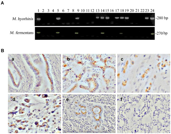
Figure 1.
Mycoplasma infection in gastric cancer tissues. A. Nested PCR of mycoplasmal DNA amplified from human gastric cancer tissues. Universal primers were used for the first round PCR and Mycoplasma hyorhinis-specific primers or Mycoplasma fermentans-specific primers for second round PCR to amplify the mycoplasmal DNA. Lanes 1 and 2 are positive and negative controls, respectively. Lanes 5, 9, 14, 18, and 24 show results from tissues from which both Mycoplasma hyorhinis and Mycoplasma fermentans DNA were amplified. Lanes 13, 15, 17, 19, and 23 show results from tissues from which only Mycoplasma hyorhinis DNA was amplified. B. IHC staining of gastric cancer tissues with a pan-specific anti-mycoplasma antibody (400 ×). (a, b, c) well-differentiated, moderately differentiated and poorly differentiated adenocarcinomas, respectively. (d) mucous adenocarcinoma. (e) lymph node metastasis of moderately differentiated adenocarcinoma. (f) control staining with IgG.

