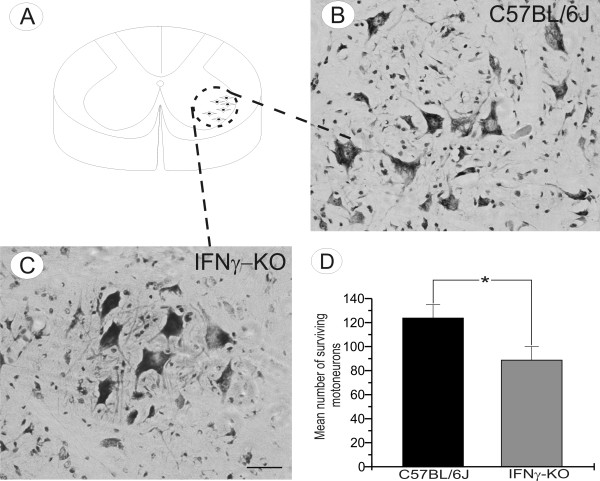
Figure 7.
Motoneuron degeneration in IFN-KO mice. (A) The schematic drawing of spinal cord section at the lumbar level demonstrating the sciatic motoneurons pool. (B-C) Representative images showing motoneuron cell bodies in unlesioned C57BL/6J and IFNγ-KO animals. (D) Graphical representation of spinal motoneuron survival. Note a significant reduction in number of motoneurons in IFNγ-KO mice. Scale bar 50 μm

