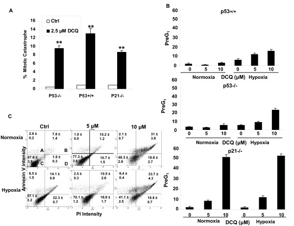
Figure 3.
DCQ induces mitotic catastrophe and apoptosis in HCT116 cells. (A) Low concentrations of DCQ triggered mitotic catastrophe in all HCT116 cell lines. Cells were cultured on coverslips and treated at 50% confluency with 2.5 μM DCQ for 48 hrs after which they were fixed and stained with Hoechst and viewed under a fluorescent microscope using UV. (**) indicates p < 0.001 (one way ANOVA) with respect to the Ctrl. (B) Higher concentrations of DCQ (5 and 10 μM) induced increases in the PreG1 phase population more so under hypoxia. Treatment with DCQ in normoxia or hypoxia was for 6 hrs, after which cells were harvested immediately and DNA was stained with PI for analysis with FACScan flow cytometry. The percentage of Pre G1 cells was calculated using Cell Quest. (C) Annexin V assay showing the apoptotic/necrotic response of p53+/+ cells exposed to 5 or 10 μM DCQ for 6 hr in normoxia or hypoxia. Apoptosis was assayed 24 hr after drug treatment, and appeared to be enhanced in hypoxia at higher drug concentrations. Quadrant A = apoptotic cells, B = apoptotic+necrotic, C = normal, D = necrotic. The experiment was repeated twice each in duplicates.

