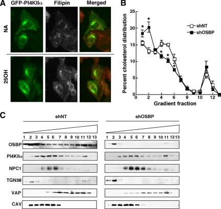
Figure 5.
OSBP depletion reduces the cholesterol content of Golgi/endosomal membranes containing PI4KIIα. (A) The localization of cholesterol was determined by filipin staining of CHO cells transiently expressing PI4KIIα-GFP and treated with or without 25-hydroxycholesterol (2.5 μg/ml) for 60 min. (B) Equivalent amounts (2–2.5 mg protein) of the postnuclear supernatants from CHO cells expressing shOSBP or a nontargeting control (shNT) were fractionated on an Opti-Prep gradient and assayed for unesterified cholesterol content as described in Materials and Methods. The cholesterol content of individual fraction is expressed as a percentage of the total cholesterol recovered from the gradient and is the mean and SEM of three experiments (*p < 0.05). (C) Distribution of the organelle markers NPC1 (late endosomes), TGN38 (Golgi), VAP (ER), and caveolin (plasma membrane/endosomes), as well as OSBP and PI4KIIα, were determined by immunoblotting of equivalent volumes of each fraction.

