Figure 3.
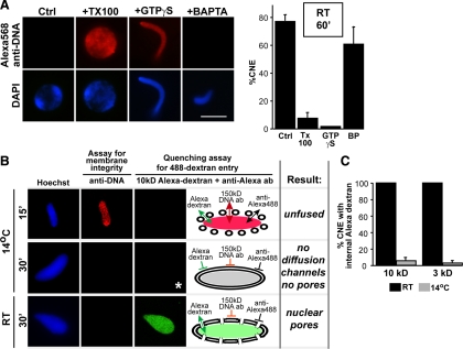
The 30-min cold intermediate containing POM121 and the Nup107-160 complex lacks diffusion channels. (A) An anti-DNA antibody exclusion assay was used to assess the formation of completely enclosed nuclear envelopes. Nuclei were assembled at room temperature for 60 min with chromatin, membranes, and either control cytosol, GTPγS-treated cytosol, or BAPTA-treated cytosol. Control nuclei were either visualized alone (Ctrl) or permeabilized with Triton X-100 (TX100) to allow entry of anti-DNA antibody. Positive labeling of the chromatin with Alexa568-anti-DNA antibody in the GTPγS nuclei indicated that a fully closed NE did not form under this condition; absence of anti-DNA antibody staining for Control and BAPTA nuclei indicated that a fully closed NE formed in these conditions. Scale bar, 10 μm. The percentage of nuclei showing a lack of DNA antibody staining in their nuclear interior (% CNE) was quantified for ∼100 randomly chosen nuclear intermediates each in three independent experiments, as shown in the histogram. Error bars indicate SD. (B) The anti-Alexa488 quenching assay described in Supplemental Figure 4 was used to determine the presence of 10-kDa competent diffusion channels in closed nuclear envelopes reconstituted for 15 min and 30 min at 14°C. The 15 min, 14°C intermediates were not fully membrane enclosed, as they showed anti-DNA staining (red) and quenching by anti-Alexa488 antibody (black) (top panels). By 30 min at 14°C, many intact nuclei had formed, as judged by a lack of DNA staining, but these showed no entry of Alexa488-10 kDa dextran indicating no diffusion channels (middle panels). In contrast, by 30 min at room temperature, the nuclei were intact (no anti-DNA staining) but had developed nuclear pores capable of allowing Alexa488-dextran entry, enabling its protection from the quenching antibody (lower panels). (C) The percentage of 14°C 30 min enclosed nuclear intermediates (i.e., with no anti-DNA staining) that showed diffusion of Alexa488-10 kDa or Alexa488-3 kDa dextrans was measured in three different experiments each and plotted. We found that the great majority of enclosed 14°C 30 min nuclear intermediates did not show diffusion channels. Errors bars indicate the SD.

