Figure 4.
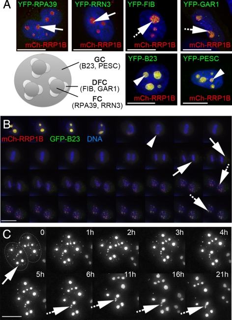
RRP1B is a GC protein that colocalizes with B23 throughout the cell cycle. (A) Coexpression of mCh-RRP1B (red) with markers (green) for the three subnucleolar compartments, the fibrillar centre (FC), dense fibrillar component (DFC), and granular component (GC), reveals that the subnucleolar distribution of RRP1B is spatially distinct from both the RPA39 subunit of RNA Pol I and its associated RRN3 factor (FC; arrows) and from Fibrillarin and Gar1 (DFC; hashed arrows). RRP1B colocalizes with B23 and Pescadillo, suggesting that it is predominantly a GC protein (arrowheads). (B) Time-lapse imaging of mCh-RRP1B (red) coexpressed with GFP-B23 (green) in U2OS cells stained with Hoechst 33342 to visualize DNA (blue) reveals that these proteins colocalize throughout the cell cycle. When nucleoli break down in prometaphase they both accumulate in the perichromatin region (arrowhead) and later appear in prenucleolar bodies (PNBs; arrows), eventually relocalizing to reformed mature nucleoli (hashed arrows). Images are 2D projections of 3D datasets captured every 10 min. (C) Time-lapse imaging of GFP-RRP1B in U2OSGFP-RRP1B cells shows the transfer of RRP1B from PNBs to nucleoli in late telophase. For most PNBs (arrow) the RRP1B signal disappears and reappears in nucleoli, although fusion of small foci was also observed (hashed arrows). Images are 2D projections of 3D datasets and time (in hours) is indicated. The two adjacent nuclei are outlined in the first panel for clarity. Scale bars are 15 μm.

