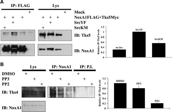
Figure 1.
The interaction between NoxA1 and Tks proteins is dependent on Src activity. (A) The interaction between NoxA1 and Tks5 is dependent on Src activity. In the left panel, HEK293 cells were transfected as indicated with Myc-tagged Tks5 and with Flag-tagged NoxA1 in presence of constitutive active or dominant negative Src (SrcYF or SrcKM respectively). After 24 h, cells were lysed and immunoprecipitation (IP) was carried out (see Material and Methods) using Flag antibody. Lys indicates lysate before IP was performed. The interaction between NoxA1 and Tks5 was tested by immunoblot (IB) using Tks5 antibody (upper section), while comparable expression levels of transfected Flag-tagged NoxA1 in cell lysates and immunoprecipitation efficiency was assessed by reblotting the membrane with NoxA1 specific antibody (lower section). One representative experiment from three separate experiments is shown. In the right panel, the results from three independent experiments performed as above were quantified. The levels of immunoprecipitated Tks5 (IP-Tks5) for each condition were normalized to the respective total Tks5 present in the Lys. The IP-Tks5 levels in presence of SrcYF were set to 1 (mean ± SEM from three independent experiments). (B) The interaction between NoxA1 and Tks4 is dependent on Src activity. In the left panel, DLD1 cells were treated as indicated with DMSO control, Src inhibitor PP2, or its nonfunctional analog PP3. After 16 h, cells were lysed and IP was carried out using NoxA1-specific antibody or pre-immune serum (P.I.). Specific interaction between endogenous Tks4 and NoxA1 was detected using Tks4-specific antibody (upper section), while the presence of NoxA1 in cell lysates was verified by reblotting the membrane with NoxA1-specific antibody (lower section). One representative experiment from three separate experiments is shown. In the right panel, the results from three independent experiments performed as above were quantified. The levels of immunoprecipitated Tks4 (IP-Tks4) for each condition were normalized to the respective total Tks4 present in the Lys. The IP-Tks4 levels in DMSO treatment condition were set to 1 (mean ± SEM from three independent experiments).

