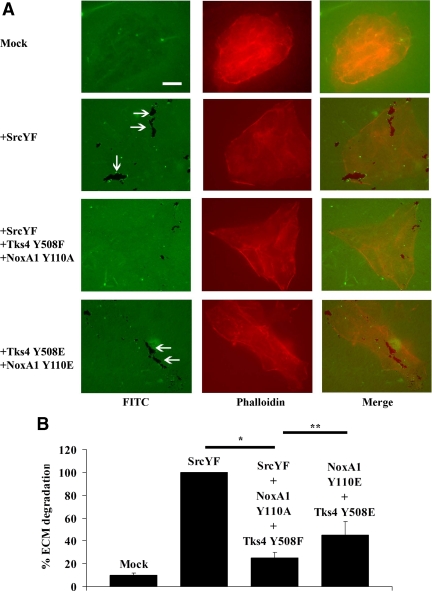
Figure 7.
The presence of Tks4 and NoxA1 unphoshorylable mutants blocks SrcYF-induced ECM degradation in DLD1 cells, whereas the presence of their phosphomimetic mutants partially rescues this phenotype. (A) The analysis of the ability of DLD1 cells to degrade the ECM shows that the presence of Tks4 and NoxA1 unphoshorylable mutants blocks SrcYF-induced ECM degradation in DLD1 cells, whereas the presence of their phosphomimetic mutants partially rescues this phenotype. DLD1 cells were transfected as indicated, and 24 h later they were trypsinized and plated on FITC-labeled gelatin-coated coverslips. After 48 h, cells were fixed in 4% PFA, stained with Alexa-Fluor-568 phalloidin, and visualized by epifluorescence microscopy (×60). The white arrows indicate areas in which cells (in red) degrade the ECM (in green). The merge is shown in the right column. Scale bars, 10 μm. One representative image from three separate experiments is shown. (B) Quantification from three independent biological experiments shown in A is given: for each experiment, the total degradation area was obtained as sum of degradation areas calculated using Metamorph software from 25 random fields and reported as percentage (SrcYF was set as 100%). In the graph, error bars represent SEM. *p < 0.01; **p <0.05 (Mann–Whitney U test).

