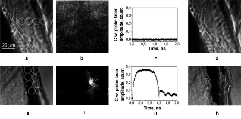
Figure 6.
(a) and (e) Bright field microscopy images of a zebrafish embryo show muscle and blood vessel areas, white circles show the areas of application of the pump laser pulses; (b) and (f) time-resolved scattering differential images of the same area obtained with the 10 ns delayed (relative to the pump laser pulse) pulsed probe laser (690 nm, 0.5 ns), white circle shows the aperture of the pump laser beam at the fluence 500 mJ cm−2 (b) and 740 mJ cm−2 (f); (c) and (g) corresponding time responses obtained during the exposure to the pump laser pulses show no bubble (c) and the bubble expansion, collapse, oscillations and residual heating after the bubble collapse (g); (d) and (h) bright field microscopy images of the same areas as in (a) and (e) obtained after their exposure to the pump laser pulses show no damage at 500 mJ cm−2 (d) and tissue disruption at 740 mJ cm−2, white circles show the point of application of the pump laser.

