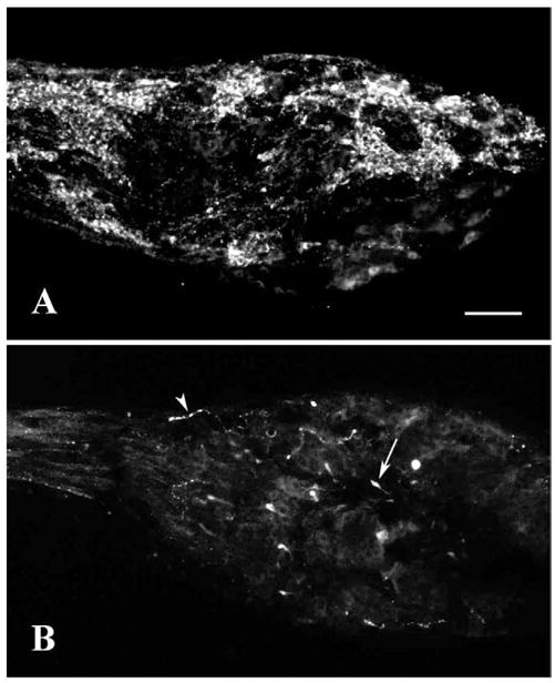
Figure 1.

Examples of differences in GnRH immunocytochemistry in the ganglion of bonnethead NT. A: Labeling with GF-5 antiserum showing large clusters of GnRH-ir cells and tracts of fibers. B: Labeling with 7CR-10 antiserum showing relatively sparse labeling of varicose fibers (arrowhead) and a few club-like structures (arrow). Scale bar = 100 μm.
