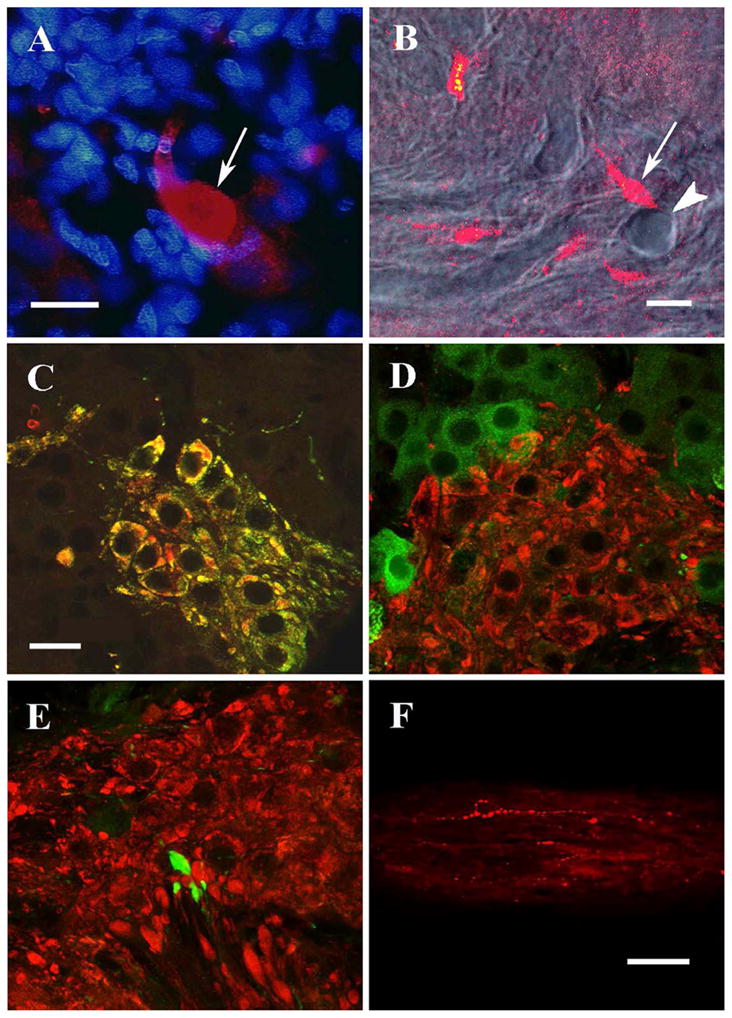
Figure 2.

Confocal micrographs of GnRH and ChAT immunocytochemistry in NT ganglion. A: Large club-like structure labeled with 7CR-10 (arrow) contains no blue-stained nuclear material (Hoechst dye). B: Combined DIC/single-confocal slice image: A club-like structure (arrow) is adjacent to a blood vessel (arrowhead) that traverses the plane of section, exiting the section near the club. C: A projected stack of confocal images showing ChAT-ir (red) and GnRH/GF-5-ir (green) are co-localized resulting in clusters of cells labeled yellow. All single confocal images also showed colocalization in all cells. D: FMFRa-ir (green) and ChAT-ir (red) are not co-localized. E: Club-like structures labeled with 7CR-10 (green) are not co-localized with ChAT-ir (red). Small areas of yellow in D and E are an artifact of stacked confocal images from adjacent optical sections superimposed on one another, and thus do not indicate co-localization. F: Central nerve trunk labeled with 7CR-10. Scale bars = 20 μm for photos A-E (D and E taken at same magnification as C). Scale bar = 50 μm for photo F.
