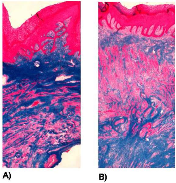
Figure 1.
Trichrome images of nulliparous (A) and parous (B) NHP. Stained bright pink is the epithelium layer, followed by the sub-epithelium layer (blue), which contains a majority of the collagen matrix of the vaginal cross-section. The muscularis bilayer consists of a layer of circumferentially oriented and a layer of longitudinally oriented muscle fibers, which are stained pink. The adventitia, which is a loose connective tissue layer stained blue.

