
FIGURE 1.
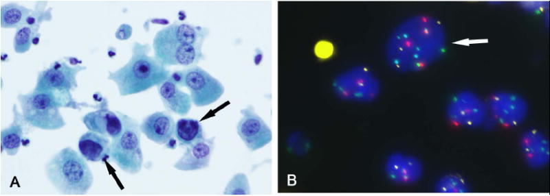
(A) A bladder washing specimen had rare atypical cells (arrow) with hyperchromatic irregular nuclei (Papanicolaou stain). (B) Atypical cell (arrow) with polysomies for chromosomes 3 (red), 7 (green), and 17 (aqua) compared to the other cells that are diploid (FISH). In this case, 6 of 25 cells analyzed showed an abnormal pattern, indicative of a positive FISH result. On clinical follow-up, the patient was found to have UC.