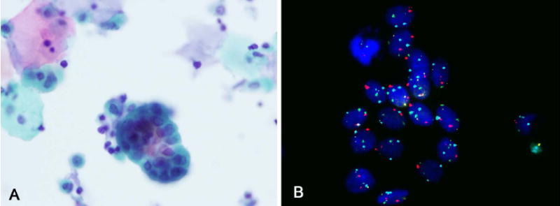
FIGURE 3.

(A) Urine cytology showed groups in a urine specimen from a patient with a history of UC (Papanicoloau stain). (B) The cells showed 2 signals for centromeric chromosomes 3, 7, 17, and locus-specific probe 9p21, indicating a negative result (FISH). On clinical follow-up the patient was negative for UC.
