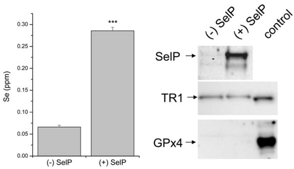
Figure 2.
Assessment of selenium and SelP content in concentrated media. Left: ICP analysis of selenium content in media samples; (+) SelP represents media from cells induced to express SelP by ponasterone A while (−) SelP represents media from cells treated with EtOH (vehicle control). The selenium difference between these concentrated media is highly significant (***, p<0.001). Right: Immunoblot analysis for SelP, TR1 and GPx4 in these media samples. The control for TR1 is 10 μg of HEK293 cell lysate, and the control for GPx4 is 30 μg of mouse testicular lysate.

