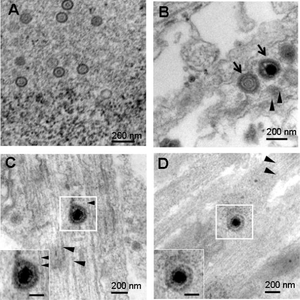
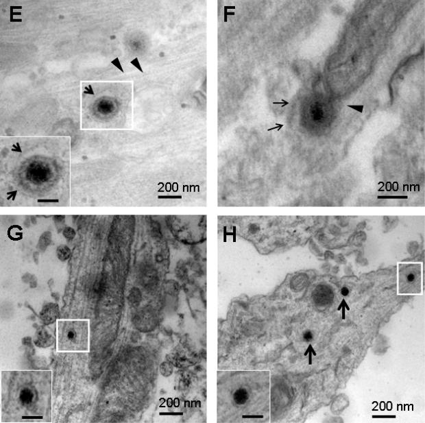
Figure 3.
Rat or mouse SCG neurons that were mock infected, or infected with 1×10⁵ PFU of HSV-1 or HSV-2 and examined by TEM at 48 hpi. (A) Nucleocapsids ~ 110 nm were detected in rat SCG nuclei after infection with HSV-1 NS. (B) Multiple virions (arrows) measuring 220 to 225 nm possibly within a large vesicle were detected adjacent to microtubules (arrowheads) in a proximal axon (S chamber) of a rat SCG neuron after infection with HSV-1 NS. (C) A virion measuring 235 nm was detected in the mid-axon of a mouse SCG neuron adjacent to microtubules after infection with HSV-1 NS. (D, E) Virions measuring 213 nm and 220 nm within vesicles (arrows) were detected adjacent to microtubules (arrowheads) in rat SCG mid-axons (M chamber) after infection with HSV-1 17. (F) A virion measuring 255 nm in a vesicle (arrows) in close proximity to microtubules after infection with HSV-2 2.12. (G) A dense core vesicle measuring 102 nm was detected in a mid-axon (M chamber) of a mock infected rat SCG culture. (H) Three dense core vesicles 110 nm in diameter were noted in a mid-axon (M chamber, boxed particle) and growth cone (arrows). Boxed areas are enlarged in insets. Bars in insets are 100 nm.


