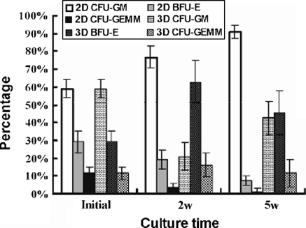
Fig. 5.
Colony distribution of the hematopoietic cells at each time point. 2 × 10⁴ UCB CD34+ cells per well were cultured in 2D or 3D system for 2–5 weeks. Cultured cells were recovered for methylcellulose CFU assay. The lineage of the colony was determined by their appearance under inverted light microscope as described in materials and methods. The percentage of each lineage was presented by mean ± SD from three separate experiments performed in duplicate. BFU-E: erythroid burst forming unit; CFU-G: granulocyte colony forming unit; CFU-GEMM: colony-forming units-granulocyte, erythroid, macrophage, megakaryocyte

