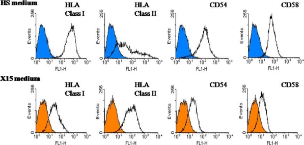
Fig. 5.
The heterogeneous expression of surface molecules per melanoma cell line established in two culture media from the same tumor-invaded LN M364. The empty histograms show staining with the mAb specific for the cell surface molecule indicated, while the overlaid filled histograms (blue for cell line established in HS medium and orange in X15 medium) represent negative control staining with isotype matched control antibody

