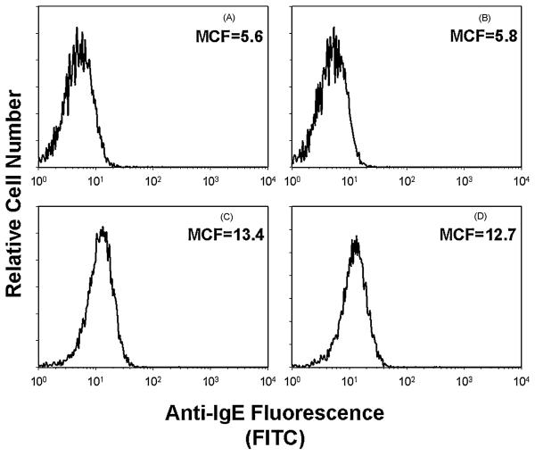
Figure 7.
Assessment of CdtBFcε for effects on IgE binding to mast cells. BMMC were incubated in medium alone (panel A), medium containing CdtBFcε (panel B; 10 μg/ml) or cells were sensitized with IgE (panels C and D) as described in Materials and Methods. The cells were then exposed to medium only (panels A-C) or CdtBFcε (panel D; 10 μg/ml) for 2 hr. The level of IgE bound to BMMC was determined by flow cytometry following staining with anti-IgE antibody conjugated to FITC. Mean channel fluorescence (MCF) is shown and indicates that similar amounts of IgE were present on BMMC in the presence or absence of chimeric toxin. It should also be noted that the anti-IgE antibody does not recognize CdtBFcε (panel B). Results are representative of three experiments.

