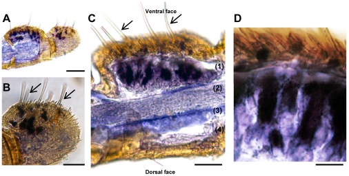
Figure 3. Expression patterns of SlCXE10 after in situ hybridization on longitudinal sections of S. littoralis male antennae.
Arrows show the olfactory sensilla. A) Global view of two antennal segments, one sectioned and the other one with intact cuticle. B) Higher magnification of A, showing labeling at the base of the olfactory sensilla. C) Longitudinal section through a segment showing the disposition of the olfactory epithelium (1), the antennal nerve (2), the antennal lumen filled with haemolymph (3) and the epidermis (4). D) Higher magnification of C showing labeled cells (accessory cells and/or neurons). Scale: 100 µm in A; 50 µm in B and C, 20 µm in D.

