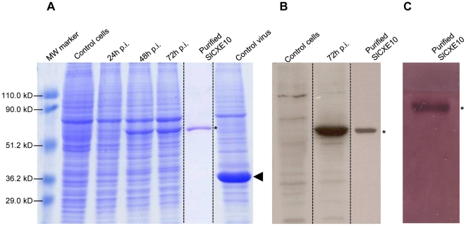
Figure 4. SlCXE10 recombinant protein expression and purification.
A) SDS PAGE analysis of total proteins extracted from infected insect cells at different times after infection (24, 48, 72 h p.i). Non-infected cells and cells infected by a non-recombinant virus were used as controls. B) Western-blot analysis. C) α/β-naphthyl acetate staining. SlCXE10 are indicated by asterisks on the right side of the gel, polyhedrin is indicated with an arrowhead. The sizes of the molecular-mass markers are shown on the left hand side of the gel.

