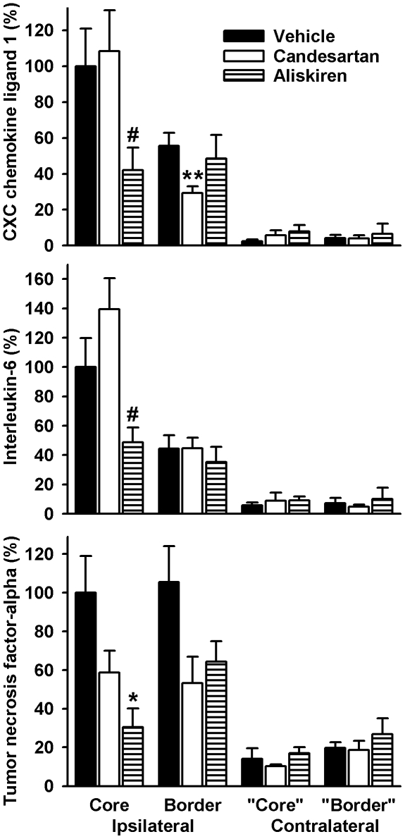
Figure 5. Relative expression of inflammatory genes in brain tissue of double transgenic rats 24 h after middle cerebral artery occlusion with reperfusion.
Tissue was obtained from the ischemic core, the border of the ischemic lesion and corresponding areas in the contralateral, non-ischemic hemisphere (“core” and “border”). Animals were pre-treated to a systolic blood pressure of 150 mmHg or received vehicle. Data are means ± standard error of the mean. n = 11-14 per group. The Mann-Whitney-U test was used for comparisons. *p<0.05, **p<0.01, vs. vehicle, #p<0.05 vs. vehicle and candesartan.

