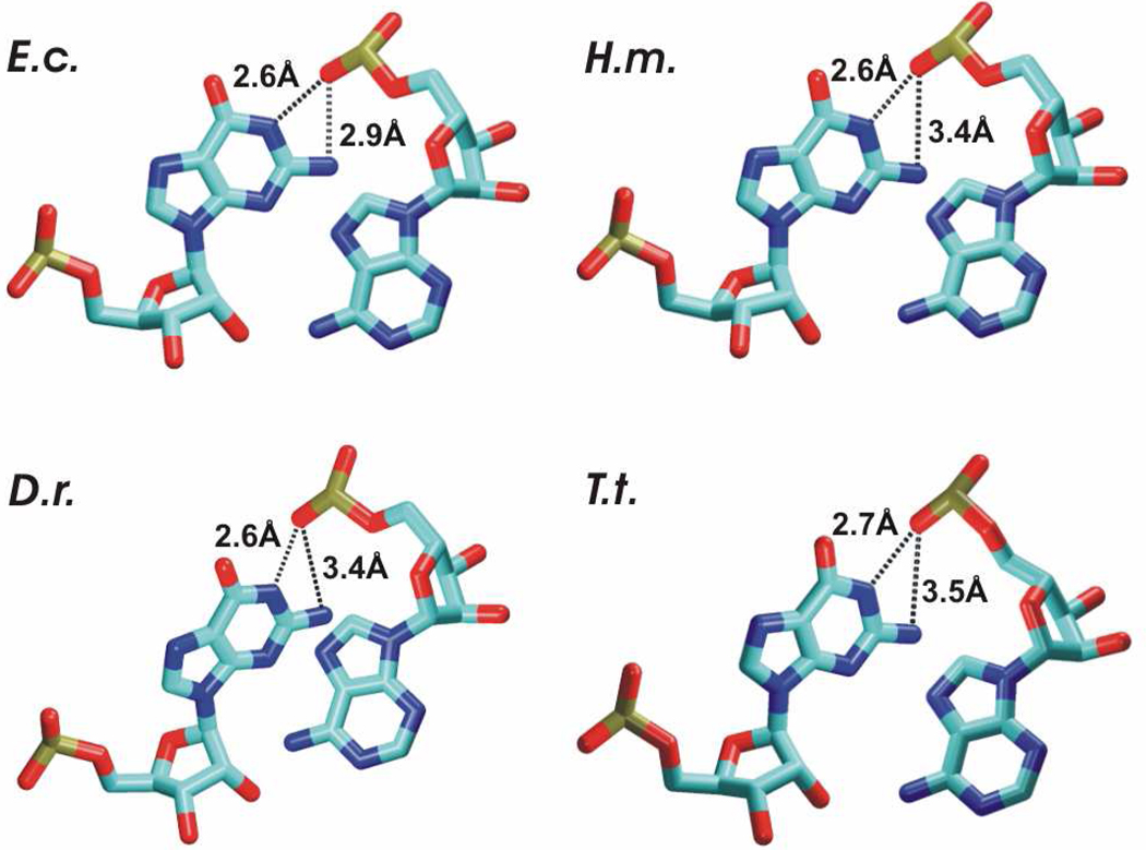
Figure 2.
Base phosphate interactions observed in the ribosomal X-ray structures of H40. E.c. structure exhibits bifurcated binding mode (base phosphate interaction type 4BPh) in which N2 and N1 of G13 bind to the same anionic oxygen of the phosphate group A5(O2P). H.m., D.r. and T.t. structures exhibit only G13(N1)-A5(O2P) H-bond, which represents base phosphate interaction type 5BPh. The differences might reflect limits of the resolution of the experimental structures.

