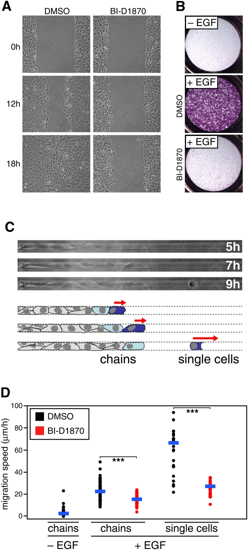
Figure 5.
RSK modulates cell migration in multiple cell migration contexts. (A) Wound scratch assay showing decreased cell migration upon addition of RSK inhibitor BI-D1870 (10 μM) in an EGF-stimulated model of cell migration using MCF10A cells. Wound scratch was introduced when MCF10A cells became confluent. Fresh media containing EGF and the inhibitor was added after introducing the wound scratch. Migration status was monitored at the indicated time points. (B) Boyden chamber assay showing decreased cell migration upon addition of RSK inhibitor BI-D1870 (10 μM) in an EGF-stimulated model of cell migration using MCF10A cells. MCF10A cells were resuspended in an EGF-free media and placed in the Boyden chamber. Where indicated, EGF was added to the media in the lower chamber. The assay duration was 24 h. (C) Schematic representation of migration phenotypic categories observed in the microfluidic device. (D) Quantitative assessment of the effects observed on cell migration in the microfluidic device. RSK inhibitor BI-D1870 (10 μM) was used. Horizontal blue bars indicate a mean value for a particular data set. Two-tailed Student's t-test was used to conduct pairwise comparisons of the data. (***) P < 1 × 10⁻⁸.

