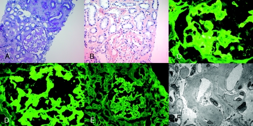
Figure 4.
Representative biopsy findings of patient 2. (A) Light microscopy showing PAS-negative amyloid material in the glomeruli, interstitium, and vessels (PAS stain, ×20). (B) Positive Congo red stain showing positive staining in the glomeruli, interstitium, and vessels (×20); immunofluorescence microscopy shows positive glomerular staining for (C) IgA and (D) κ light chains. (E) λ light chains are essentially negative. (F) Electron microscopy shows randomly oriented amyloid fibrils in the glomerulus.

