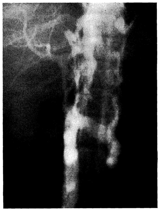
Fig. 6.

Inferior venocavagram performed 3 months after clinical portacaval transposition. The portacaval anastomosis is indicated (arrow). Note that some dye reaches the liver, but a large amount of the vena caval blood passes through paravertebral collaterals.
