
Fig. 5.
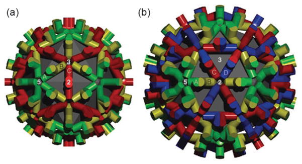
Surface lattice of T = 3 and T = 4 HBV capsids. Shown is the arrangement of dimers in the T = 3 (a) and T = 4 (b) icosahedral geometries. The quasi-equivalent sites are labelled in both cases (T = 3; A, B, C and T = 4; A, B, C, D). Note that sites with the same symbol do not have identical structures. The 5-, 3-, and 2-fold axes of symmetry are also indicated. In the T = 3 lattice, the red C dimer lies on the two-fold axis of symmetry, and across two facets of the icosahedron. No such environment exists in the T = 4 geometry. Figure reproduced from ref. 3.