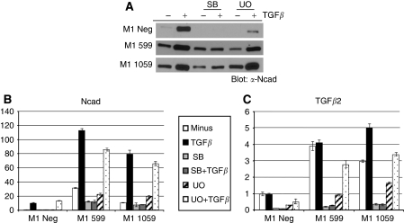
Figure 4.
Inhibition of TGFβ or MAPK signalling abrogates TGFβ-mediated EMT. (A) M1 Neg, M1 599 and M1 1059 cells were either untreated or pre-treated for 30 min with 10 μM SB431542 (SB) or 10 μM U0126 (UO) followed by addition of TGFβ (2.5 ng ml−1) for 48 h. Cell lysates were prepared and western blot analysis performed to determine N-cadherin (Ncad) expression levels as described in Materials and Methods (n=2 independent experiments). (B, C) qRT–PCR analysis was performed to determine relative expression levels of N-cadherin, TGFβ1, TGFβ2 and TGFβ3 mRNA following the treatments described in A. Shown is the mean±s.d. of triplicate assays with M1 Neg unstimulated levels set to 1.0 (n=2 independent experiments).

