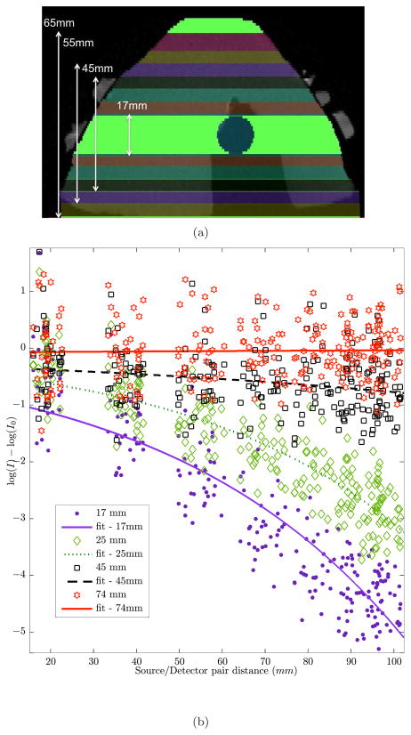
Fig. 5.
(a) Different heights from the MR image of the the breast phantom (17, 25, 35, 45, 55, 65 and 74mm) were used to create various sizes for the 3D breast mesh to study the effect of trimmed meshes. (b) Using the full breast mesh as the reference, the change in light fluence of the trimmed meshes (shown in (a)) at the detector locations is shown here. Horizontal axis is the distance between source/detector pairs and vertical axis is the light fluence difference between full mesh (I0) and partial mesh (I). For clarity not all trimmed sizes are shown. The height of the full breast mesh was 79mm.

