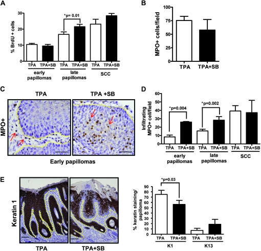
Fig. 4.
Early papillomas from SB-treated mice have a progressed phenotype. (A) Tumor cell proliferation in early (n = 6) and late papillomas (n = 10) and SCC (n = 5) measured by bromodeoxyuridine incorporation counted at magnification ×400. (B) No significant change in total neutrophils (MPO+ cells) per field, counted at magnification ×400 (n = 6). (C) Localization of neutrophils in early papillomas from TPA-and SB/TPA-treated mice at magnification ×400 with scale bar representing 20 μm. Arrows indicate neutrophils. Yellow line shows epidermal/dermal junction. (D) Effect of SB on tumor infiltrating neutrophils in early (n = 6) and late (n = 18) papillomas and SCC (n = 5). (E) Reduced keratin 1 levels in early SB-treated papillomas (left). Representative micrographs, magnification ×100, scale bar representing 50 μm. Yellow line shows epidermal/dermal junction. Decreased expression of keratin 1 and increased expression of keratin 13 messenger RNA in early papillomas treated with SB (n = 12) (right). Percent keratin staining in the skin was scored blindly by two individuals.

