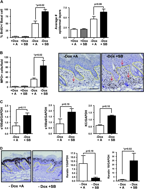
Fig. 5.
SB rapidly induces progressed phenotype in epidermis-expressing human C-Ha RASV12G oncogene. (A) Significant increase in proliferation with SB treatment in RAS-expressing skin (left) and slight increase in epidermal thickness (right). (B) An increase in MPO+ cells in the dermis of SB-treated mice expressing the InvtTA × tetORASV12G transgenes. Arrows indicate neutrophils. Yellow line shows epidermal/dermal junction. Representative micrographs (right), magnification, ×100. Scale bar represents 50 μm. (C) Increased messenger RNA expression of neutrophil chemokines s100a8, s100a9 and keratinocyte chemokine with treatment of SB. (D) SB causes significant decrease in keratin 1 staining (left) and messenger RNA expression (middle). Representative micrographs (left), magnification ×100. Scale bar represents 50 μm. Yellow dotted line shows epidermal/dermal junction. Significant increase in keratin 18 messenger RNA expression by quantitative reverse transcription–polymerase chain reaction in RAS-expressing skin following SB (right). n = 4–6 mice per treatment group.

