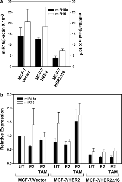
Fig. 4.
HER2Δ16 expression suppresses miR-15a and miR-16. (a) Expression of miR-15a and miR-16 is suppressed in MCF-7/HER2Δ16 cells. Total RNA was extracted and analyzed for miR-15a or miR-16 expression by qRT–PCR. Results from three independent RNA extractions are represented as mean ± SE expression relative to β-actin. The lower levels of miR-15a and miR-16 expression in the MCF-7/HER2Δ16 cells failed to reach significance (paired Student’s t-test; P = 0.07 and P = 0.08, respectively). (b) Expression of miR-15a and miR-16 is not altered by estrogen or tamoxifen. Each cell line was cultured for 48 h in phenol red-free modified Eagle;s medium containing 5% charcoal-stripped fetal bovine serum and then left untreated or treated for 16 h with 100 pM E2 alone or in combination with 1.0 μM TAM. Three independent total RNA extractions from each cell line were analyzed in triplicate for miR-15a and miR-16 expression by qRT–PCR. Results were normalized to β-actin and represented as mean ± SE expression relative to untreated MCF-7/Vector cells. Differences failed to obtain significance as determined by paired Student’s t-test.

