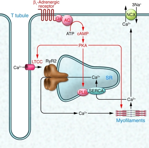
Figure 1. The classical view of cardiomyocyte excitation-contraction coupling and its regulation by β-adrenergic receptors.
Under unstimulated conditions (black arrows), depolarization during an action potential opens LTCCs in T tubules, allowing Ca2+ to enter the cell. This trigger Ca2+ induces a larger Ca2+ release from the SR via RyR2. The increase in Ca2+ concentration initiates conformational changes of the myofilaments and thereby contraction. Removal of Ca2+ via SERCA and NCX reverses the process. Catecholamines stimulate excitation-contraction coupling (red symbols and lettering) by phosphorylating LTCCs (increased Ca2+ influx), PLB (increased Ca2+ reuptake into the SR), and myofilament-based troponin I and myosin-binding protein C (increased relaxation). AC, adenylyl cyclise; Gs, stimulatory G protein. Adapted with permission from Nature (35).

