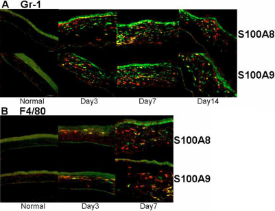
Figure 2.
Immunostaining for S100A8, A9, and cellular markers in murine corneas. Neutrophils and macrophages were stained green via primary rabbit anti-Gr-1 (A) or anti-F4/80 (B) and FITC-conjugated secondary antibodies, while S100A8 or A9 were stained red via PE conjugated primary antibodies. Please note that the autofluorescence for corneal epithelium should not be misinterpreted as positive staining.

