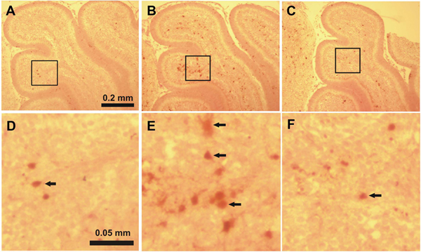
Figure 2.
Immunostaining for activated caspase-3 in lobule IV-V of the cerebellum of 7-day-old mice 8 hours after the first ethanol injection A, D: control group, B, E: ethanol-treated group, C, F: ethanol+taurine-treated group. The activated caspase-3-immunoreactive cells in the IGL are indicated by black arrows.

