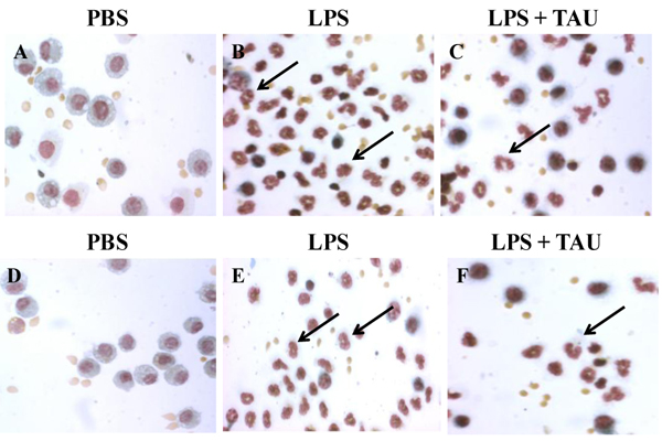
Figure 9.

Photomicrographs showing cells in BALF samples after a staining with Wright’s solution. The animals received TAU (50 mg/kg/0.5 mL, i.p.) before (A-C) and after (D-F) LPS (0.02 mg). Cells from control (PBS pH 7.4) animals exhibited a normal differential count, with the majority of cells being macrophages (A and D). BALF from animals treated only with LPS (B and E) exhibited a higher number of neutrophils and only a few macrophages relative to BALF from control (PBS pH 7.4) animals. A 3-day treatment with TAU, either before (C) or after (F) LPS, reduced the number of neutrophils relative to BALF from animals receiving only LPS (magnification of 400x).
