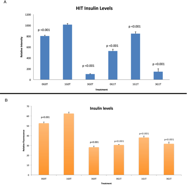
Figure 3.
Comparison of insulin levels in Hit and Rin cells. Relative fluorescence intensity, a marker for amounts of protein present in the cell, from cells represented in Figures 1 and 2 For statistical analyses, all p values are related to control cultures treated with 1 mM glucose only. A: Relative fluorescence intensity for insulin in the β-cell line Hit-T15. B: Relative fluorescence intensity for insulin in the β-cell line Rin-m. Using 1 mM glucose in the absence of taurine (1G0T) as a control, all treatments resulted in significantly reduced insulin immunoreactivity. Legend: 0G0T (no glucose or taurine), 3G0T (3 mM glucose, no taurine), 0G1T (no glucose, 1 mM taurine), 1G1T (1 mM each glucose plus taurine), 3G1T (3 mM glucose, 1 mM taurine).

