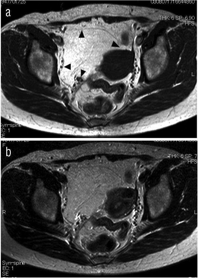
Figure 1.

Retropelvic mass showed very high intensities on T1-(a) and T2-(b) weighted images that were the same intensity as fatty tissues. This mass was diagnosed as a retropelvic lipoma by observing the position of the uterine broad ligament (arrow), the round ligament (arrow), the uterine artery (arrow) and the lateral umbilical ligament (arrow).
