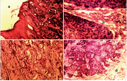
Figure 1.
a, b, c, d. Histology of the various sections of the tumor; (a) Normal skin and contiguous areas the well differentiated squamous cell carcinoma invading the surrounding deeper tissue; (b) High magnification of the malignant squamous cells; (c) Remnants of the testicular tubules being invaded by the tumor; (d) The malignant squamous cells invading the deeper testes.

