
Figure 4.
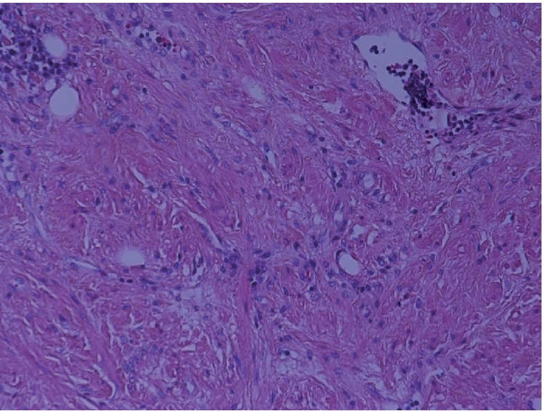
Microphotograph showing smooth muscle fibres containing benign spindle cells, together with variably sized blood vessels in a background of fibrous tissue (H&E ×200).
Official websites use .gov
A
.gov website belongs to an official
government organization in the United States.
Secure .gov websites use HTTPS
A lock (
) or https:// means you've safely
connected to the .gov website. Share sensitive
information only on official, secure websites.
Microphotograph showing smooth muscle fibres containing benign spindle cells, together with variably sized blood vessels in a background of fibrous tissue (H&E ×200).